
What Does Fourier Transform Do to Cryo-EM Images?
Published:
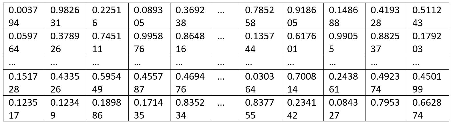
Cryo-EM images are formed by pixels with different intensity in grayscale. Each pixel is represented as a number in the image file. Below is a table simulating the pixel intensity of an image.
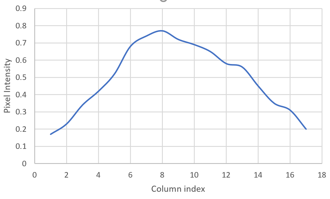
If we plot the numbers in the last row with column indexes, we may get a curve look like this:
Here we need to introduce a theorem that every curve can be approximated by the sum of simple sine waves (y = A sin(wx +b)) as presented in the figure below. Therefore, if we apply Fourier transform to the curve, we will get a set of sine waves with different amplitude (A), phase (b) and frequency (1/w). In this way, we convert the information in the real image into three parameters in the Fourier space. And the process of converting a curve into several sine waves is called Fourier transform.
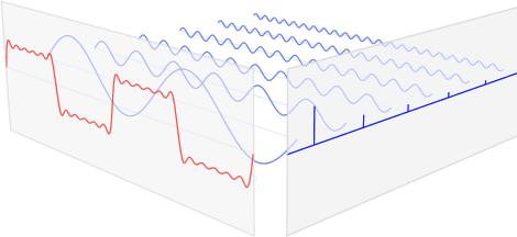
The red curve on the left is able to be approximated by the sum of several blue curves on the right. Image from https://www.ritchievink.com/blog/2017/04/23/understanding-the-fourier-transform-by-example/
In the Fourier transformed image, we record two numbers, amplitude and phase in the order of frequency from low to high. For example, if we have a real image with image size 100x100 and pixel size=2 Å, the Fourier transform of one row in the image would be 100/2=50 sine waves ordered from 0 frequency to the Nyquist frequency which is 0.25 Å-1 (1/(pixel size*2)).